Why Did Sabertooth Tigers Need Such Big Teeth? UB Team "Retro-Engineers" Ancient Beasts to Find Answer
Fields that can benefit range from evolutionary biology to medicine
Release Date: January 8, 2004 This content is archived.


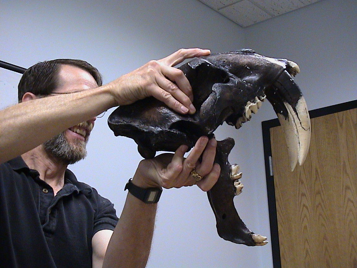
BUFFALO, N.Y. -- Cringe. That's what most people do when they look at fossils of the impressive, eight-inch-long canines of the now extinct sabertooth tiger, Smilodon fatalis.
But Frank Mendel, a University at Buffalo anatomist, sees those big teeth and thinks: How in the world did they use those fangs?
A team of design engineers in UB's New York State Center for Engineering Design and Industrial Innovation (NYSCEDII) is developing the first interactive, computational toolkit designed to answer Mendel's question -- and others like it concerning how ancient beasts behaved -- with mathematical precision.
"We are creating a computer-aided design (CAD) system for living things," said Mendel, Ph.D., associate professor of pathology and anatomical sciences in UB's School of Medicine and Biomedical Sciences.
CAD allows engineers to develop three-dimensional models of buildings, automobiles, airplanes or other complex systems on computers, but until now it hasn't been applied to living organisms.
The objective of the Vertebrate Analyzer, as the CAD system is called, is to create, mechanically articulate and animate skeletal models, "fleshing" them with anatomically and physiologically correct "virtual tissues" that respond according to their own biomechanical capacities and limitations.
"The hope is to build very accurate models so that we can better understand the relationships among form (anatomy), function (physiology) and behavior," explained Mendel.
Ultimately, UB researchers say, the Vertebrate Analyzer will be able to simulate accurately and comprehensively virtual models of any vertebrate species, modern or extinct, and to demonstrate whether or not certain hypothetical behaviors are or were mechanically possible.
"The point of the toolkit is to be able to experiment with the form and function of animals," explained Kevin Hulme, Ph.D., NYSCEDII's research associate for engineering design and lead visualization scientist of the center's Vertebrate Analyzer research team. "We want to provide users with the freedom to add their own functions and modify existing features."
That versatility, they say, eventually could have significant potential for medical and dental applications. From paleontologists who want to build a virtual jaw of Tyrannosaurus rex to doctors who want to study what happens to bones and ligaments in the human knee or to the TMJ (temporomandibular) joint after injury, the ultimate potential of the Vertebrate Analyzer is enormous, its creators say.
For evolutionary biologists, the Vertebrate Analyzer is designed to allow them to do what has never before been possible: to "experiment" with extinct mammals, from sabertooth cats to dinosaurs, potentially solving some of the biggest questions remaining about ancient beasts.
And while other computational methods of reconstruction may have more aesthetic appeal, team members explained that the Vertebrate Analyzer will answer those questions with far more precision, mathematically.
"We have built into the analyzer the basics about the geometry of muscles," said Hulme. "This allows the researcher to vary the properties of a digital muscle, such as its diameter, its unstretched length, its physical and material properties, and the number of fibers (sarcomeres) in the muscle. So other types of reconstructions might be more aesthetically pleasing, but they don't have the science to support, for example, the jaw movements or biting motions they simulate."
"One of the great things about the VA is its potential to emulate almost anything," added Mendel. "We expect it to be capable of helping researchers determine, for example, if the T. rex could, indeed, have bitten through the armor plate of a duckbill dinosaur. We have chunks of duckbill skin, so we should be able to characterize it and see if the teeth of T. rex could have penetrated it without breaking."
To test Mendel's own hypothesis that Smilodon's fangs were used to cut the throats of prey, rather than suffocate them as modern cats do, the Vertebrate Analyzer team first must move briefly out of the virtual world and into the real world.
This spring, members of the VA team with expertise in robotics will build mechanical models of Smilodon and modern-day tiger skulls, complete with hydraulic jaws and fangs of aluminum or dental materials in order to choreograph attacks on horse, bison or cow carcasses.
"Using parts of fresh carcasses, which we will buy from local butchers, we can recreate the biting process and calibrate our virtual models with real numbers," explained Mendel.
Once the biting experiment is done, the carcasses will be dissected, providing the UB team with the precise data on the minimum forces the Smilodon would have had to generate to overcome the resistance offered by the tissues of the prey's neck.
So far, the group has assembled virtual models of the jaws and skulls of a human, a lion, a tiger and a Smilodon.
The model of the Smilodon skull is based on CT images of a fossil taken at Veteran's Hospital in Buffalo.
Using numerous off-the-shelf software packages along with some programs they wrote themselves, Hulme and colleagues at NYSCEDII converted those two-dimensional images into accurate, three-dimensional digital models.
At that point, the digital data then were "loaded" into the Vertebrate Analyzer.
The user then inputs information on the geometry of the muscles involved, such as their length and diameter, as well as the physical structure of the muscles and fibers, such as limits on tension and compression, if known, and material strength.
"When this step is complete, the user simply points and clicks on the model to attach the muscles to it," explained Hulme, "allowing the user to perform many trial scenarios quickly and easily."
The VA toolkit allows users to navigate (i.e., rotate, translate and zoom) around the model with a conventional PC mouse and keyboard. A perimeter display window shows data on model fidelity, jaw angle, collision properties and attached muscle characteristics.
In addition to Mendel and Hulme, the UB team developing the Vertebrate Analyzer includes Kevin Chugh, Ph.D., NYSCEDII research associate for visualization, Venkat Krovi, Ph.D., assistant professor of mechanical and aerospace engineering, David R. Pendergast, Ph.D., professor of physiology and biophysics, Abani Patra, Ph.D., associate professor of mechanical and aerospace engineering, and Scott H. Woodward, director of engineering design services for the UB School of Engineering and Applied Sciences.
NYSCEDII, based in the UB School of Engineering and Applied Sciences, was established as a major center for scientific visualization and virtual reality to serve the academic community and provide companies throughout New York State with a significant competitive advantage in high-tech product development. It provides basic research, education and training, and industrial outreach in immersive and high-end visualization, rapid virtual prototyping, Internet-based systems for design, computer-assisted design graphics and three-dimensional modeling, real-time interactions with design and analysis simulations, visual interaction with high-performance computing applications, sensory and haptic tools and interactions with virtual simulations.
Media Contact Information
Ellen Goldbaum
News Content Manager
Medicine
Tel: 716-645-4605
goldbaum@buffalo.edu