Using laser ‘tweezers,’ scientists grab and study tiny protein droplets
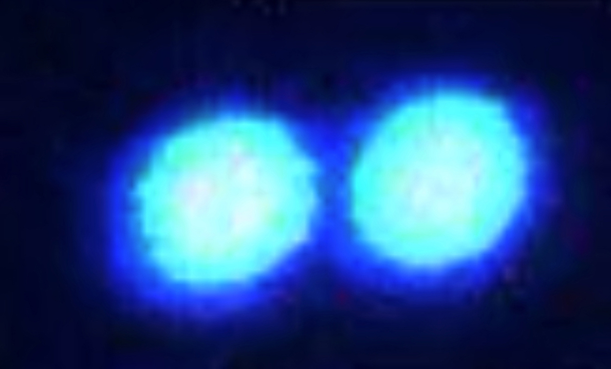
Using laser “tweezers,” University at Buffalo scientists grab protein droplets and push them together — experiments that reveal information about how hard or fluid the droplets are. The white scale bar in the top-right corner represents a length of 5 microns.
Release Date: March 7, 2019 This content is archived.


BUFFALO, N.Y. — University at Buffalo physicists are using innovative tools to study the properties of a bizarre class of molecules that may play a role in disease: proteins that cluster together to form spherical droplets inside human cells.
The scientists’ latest research sheds light on the conditions that drive such droplets to switch from a fluid, liquidy state to a harder, gel-like state.
Published on Feb. 19 in the journal Biomolecules as a featured article, the study finds that certain protein droplets harden, becoming gelatinous in crowded environments (such as test tubes where lots of other molecules are present, mimicking the congested conditions inside living cells).
IMAGES: https://buffalo.box.com/s/vls1unzwcq9sz9fqfhfzykvkq6voeiln
“These droplet-forming proteins are a relatively new area of study, so we know very little about their basic properties,” says lead investigator Priya R. Banerjee, PhD, assistant professor of physics in the UB College of Arts and Sciences. “As physicists, we want to quantify the dynamics of these droplets and learn what factors influence them. This is important as the dynamics of protein droplets are a key to their cellular function and dysfunction.
“Prior research has focused on the structure of the proteins themselves, but our work shows that environmental factors are equally important. We see that external conditions can alter the internal state of the droplets, which may affect their function in human cells.”


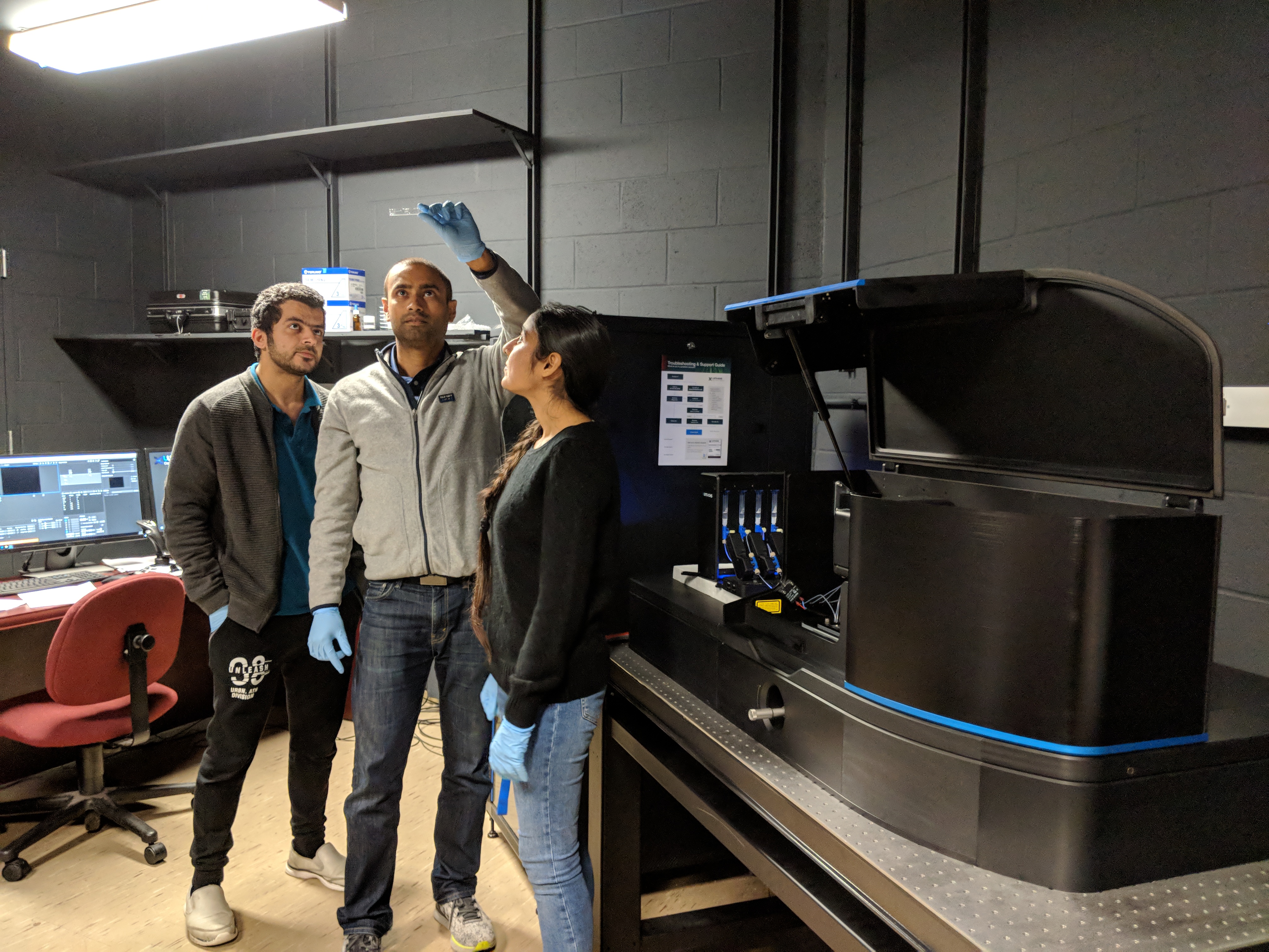
UB Assistant Professor of Physics Priya Banerjee (center) examines a microfluidic chip containing protein droplets in the lab, as UB PhD students Ibraheem Alshareedah (left) and Taranpreet Kaur (right) look on. Kaur was first author of the study, and Alshareedah was second author. Credit: Priya Banerjee Lab at UB
The research matters because condensating proteins may be involved in health and disease. Recent studies point to potential roles for these droplets in such diverse functions as gene expression, stress response and immune system function.
The new paper investigates a droplet-forming protein called fused in sarcoma (FUS). Liquid FUS droplets are found in normal brain cells, but in some patients with the neurodegenerative disease amyotrophic lateral sclerosis (ALS), the protein forms aggregates of solid material, Banerjee says. It’s unclear why.
Using lasers to tweeze and poke protein droplets


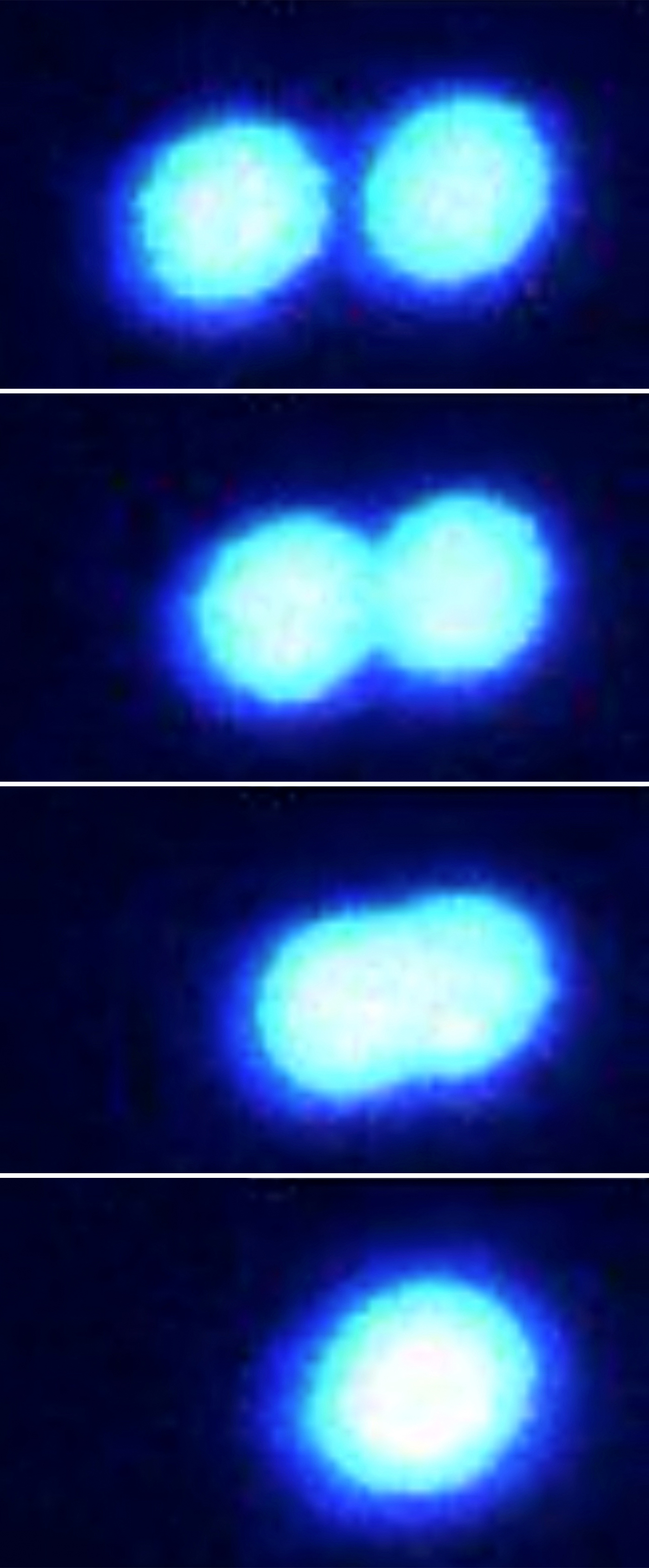
Two protein droplets fuse easily when pushed together (shown sequentially in fluorescence microscope images, top to bottom.) The droplets, made from fused in sarcoma proteins, have a fluid consistency as they sit in a solution sparsely populated by other molecules. Scientists used state-of-the-art optical tweezing technology to grab and manipulate these protein micro-droplets. Credit: Priya Banerjee Lab at UB
The research employed two innovative techniques to show how environmental conditions can affect droplets made from FUS or other related proteins.
In one set of experiments, scientists used highly focused laser beams — called optical tweezers — to trap and push together two protein droplets floating in a liquid buffer solution.
The protein droplets merged easily to form a single larger droplet when the buffer was thinly populated with other inert crowder molecules such as polyethylene glycol (PEG). But when the concentration of PEG or other chemicals in the buffer increased, the protein droplets became more gelatinous and would not fully combine.
In a second set of tests, the team employed lasers in a different way — “laser poking” — to study how FUS and related protein droplets react to crowded environments.
In these experiments, Banerjee and colleagues attached fluorescent tags to numerous protein molecules in a single droplet, causing the proteins to glow. The researchers then “poked” the middle of the droplet with a high-intensity laser, a procedure that caused any fluorescent molecules hit by the laser to go permanently dark.
Next, scientists measured how long it took for new glowing proteins to move into the darkened area. This happened quickly in protein droplets floating in sparsely populated buffer solutions. But the recovery time was dramatically slower for droplets suspended in buffer solutions thick with PEG or other compounds — an indication, once again, that protein droplets become gelatinous in crowded environments. The findings applied to both FUS and other related protein droplets with diverse primary structures.
“Our experiments were done in test tubes, but our results suggest that inside living cells, the crowding status could affect the dynamics of protein droplets,” Banerjee says.


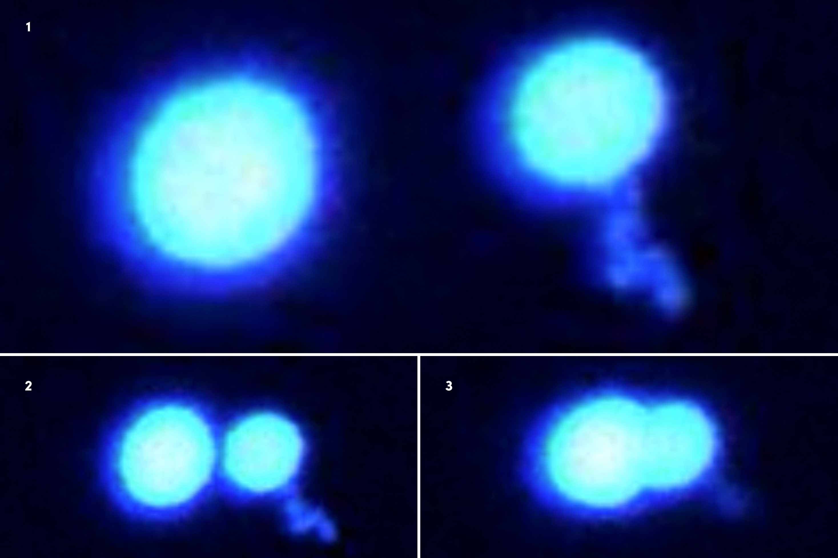
Two protein droplets refuse to merge when pushed together (fluorescence microscope images 1-3, shown sequentially). The droplets, made from fused in sarcoma proteins, are in a hard, gel-like state as they sit in a solution crowded with other molecules. Scientists used state-of-the-art optical tweezing technology to grab and manipulate these protein micro-droplets. Credit: Priya Banerjee Lab at UB
One important question that remains is whether and how the fluidity of FUS droplets impacts the protein’s ability to form into solid clumps, as seen in some ALS patients. Banerjee hopes to address this problem through future research.
The study in Biomolecules was supported by the UB College of Arts and Sciences, with assistance from the UB North Campus Confocal Imaging Facility, which is supported by the National Science Foundation. The research was conducted by Banerjee’s team at UB, with technical assistance from a colleague affiliated with Baylor College of Medicine.


Two protein droplets fuse easily when pushed together (fluorescence microscope images 1-4, shown sequentially.) The droplets, made from fused in sarcoma proteins, have a fluid consistency as they sit in a solution sparsely populated by other molecules. Scientists used state-of-the-art optical tweezing technology to grab and manipulate these protein micro-droplets. Credit: Priya Banerjee Lab at UB
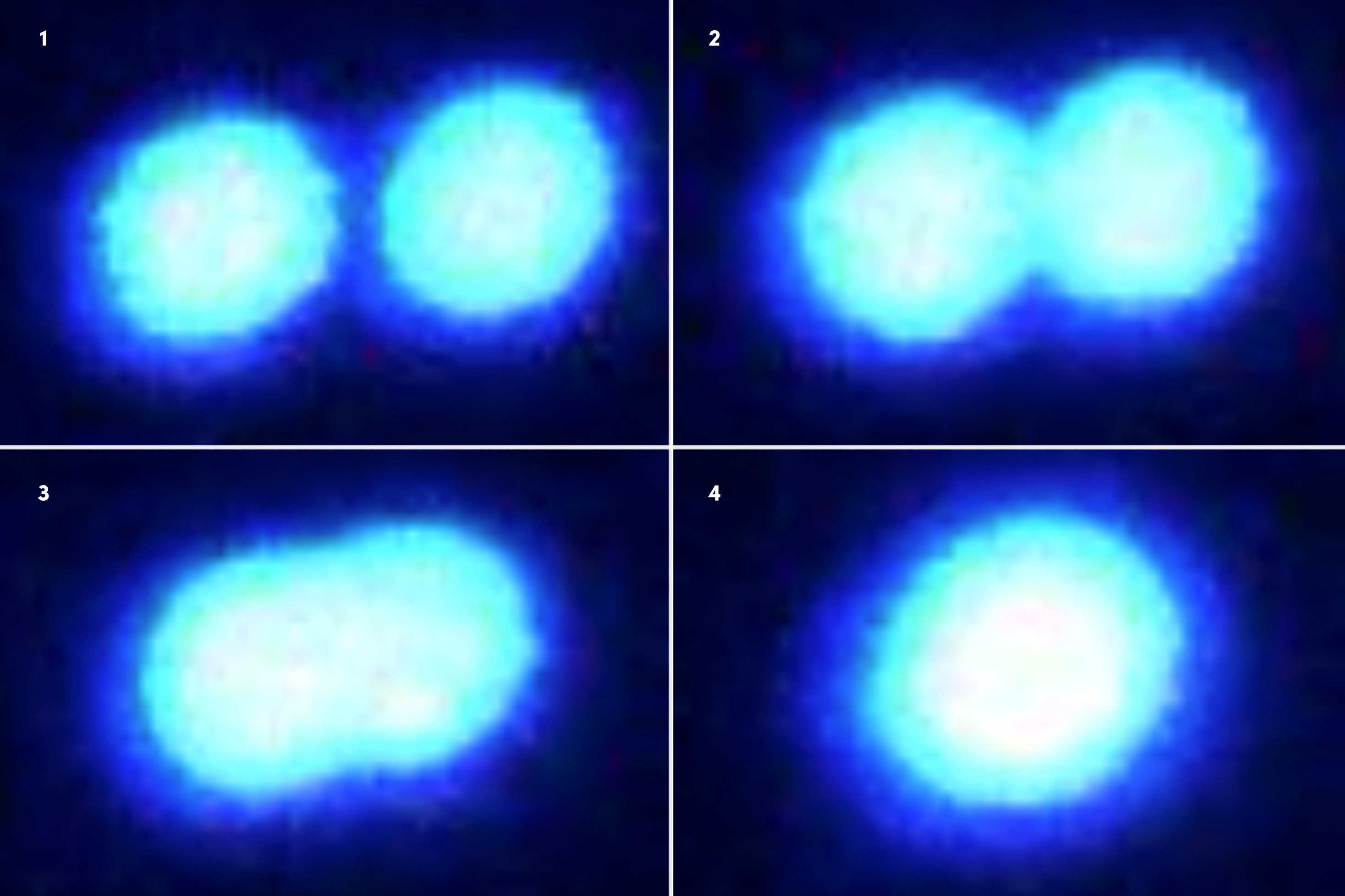
Media Contact Information
Charlotte Hsu is a former staff writer in University Communications. To contact UB's media relations staff, email ub-news@buffalo.edu or visit our list of current university media contacts.